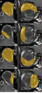
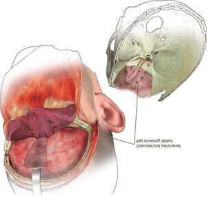
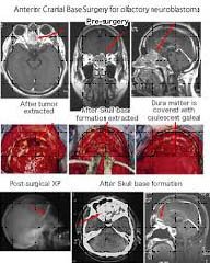
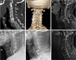
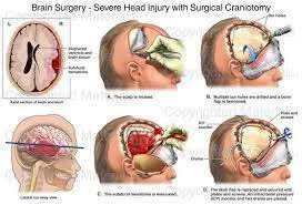
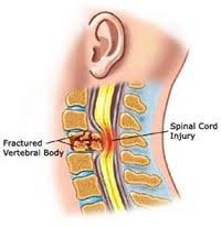
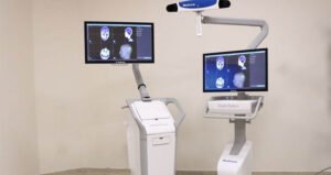
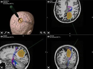
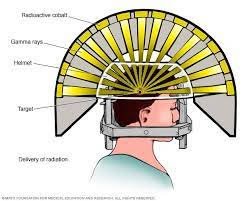
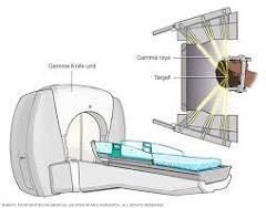
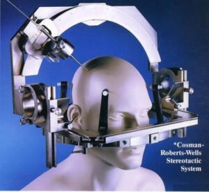
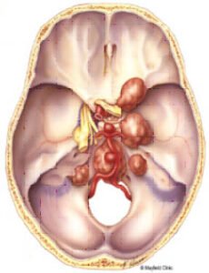
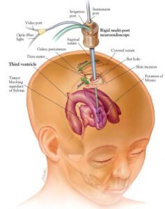
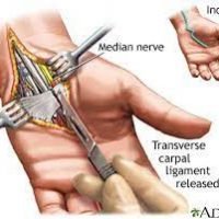
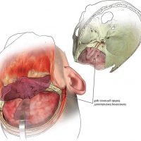
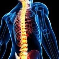
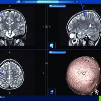
Gallery Dr. Sharadendu Narayan Detailed MRI scans illustrating different views of the knee joint. Educational illustration detailing the internal anatomy of the human head, including the brain and nasal passages. Sequential medical images documenting the progression from pre-surgery to post-surgery of a cranial base surgery. Diagnostic imaging series illustrating cervical and lumbar spine conditions. Illustrative progression of a surgical craniotomy for treating severe head trauma. Illustration of a spinal cord injury resulting from a fractured vertebral body in the cervical spine. State-of-the-art imaging equipment displaying detailed brain scans, used for diagnostic and surgical planning. Advanced brain scans and 3D modeling used for precise surgical planning of a brain tumor. Diagram illustrating the Gamma Knife procedure for precise delivery of radiation to brain tumors. Diagram showing the Gamma Knife unit and how gamma rays are precisely directed to treat brain tumors. Advanced stereotactic system for precision in neurosurgical procedures. Detailed illustration of the base of the human skull highlighting critical arteries and nerves. Detailed illustration of a neurosurgical procedure to access the third ventricle of the brain using a rigid multi-port neuroendoscope. Detailed illustration of carpal tunnel release surgery, highlighting the release of the transverse carpal ligament and the median nerve. Detailed illustration of cranial surgery dissection, highlighting the internal structures of the head and brain. Detailed illustration of the human spine within a semi-transparent body, highlighting the skeletal structure MRI series showcasing coronal, sagittal, and axial views of the brain, along with a 3D reconstruction of the skull.